OpenDerm is an open-source robotic imaging system for creating high-resolution, reproducible 3D maps of the skin. Registered scans help reveal new lesions and identify subtle changes in existing ones.
The hardware designs, software, and build documentation are all openly available.
For melanoma, early detection can make the difference between a highly treatable cancer and a life-threatening one.
More than 100,000 Americans are diagnosed with melanoma each year. This form of skin cancer has a five-year relative survival rate of nearly 100% when detected early, but about 34% after it reaches distant parts of the body.1 Approximately 30% of melanomas develop within an existing mole, while 70% appear as new lesions.2 In either case, the earliest visible sign may be small: a millimeter-scale change at the edge of a mole, a localized shift in its color or structure, or a new spot only a few millimeters across. Detecting changes at this scale requires a precise record of the skin’s prior appearance.
For people with many moles, detecting change requires tracking the entire skin surface over time. A clinician can examine the skin during an office visit, but without a standardized photographic baseline, determining whether one spot among hundreds is new or changing can be difficult. Total-body photography can provide this longitudinal record, but access to such systems remains very limited due to high costs and limited clinical evidence that existing systems reliably catch melanoma earlier than dermatologists.34
Improving total-body imaging requires progress in three areas:
01
Higher Resolution Imaging
Many existing total-body photography systems use wide fields of view to capture large areas of skin, but at the expense of fine detail. Detecting subtle changes in a lesion’s boundary, color, or internal structure requires higher-resolution images.
02
Longitudinal datasets
Most AI models for skin-cancer detection are trained on isolated images of lesions already identified as suspicious. These datasets therefore provide little evidence of how melanomas first emerge and evolve before drawing clinical attention. Training models to recognize earlier signs of melanoma will require repeated, high-resolution images of the same skin over time.
03
Frequent, accessible scanning
Total-body photography must become more affordable and widely available. Patients at high risk should be able to receive frequent, standardized scans.
A robotic imaging system offers a practical way to meet these requirements. By moving close to the body and following its contours, the system can maintain consistent distance, angle, focus, and lighting while capturing high-resolution images across the skin surface. Using a single camera with cheap actuators also reduces hardware cost compared with a large array of fixed cameras.
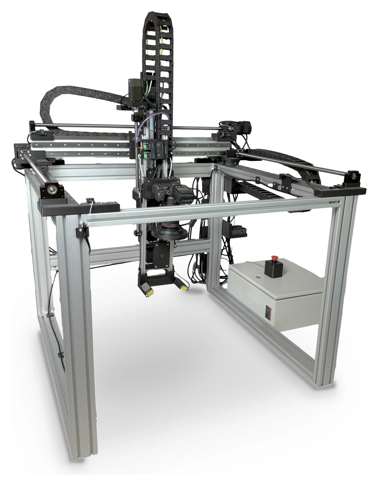
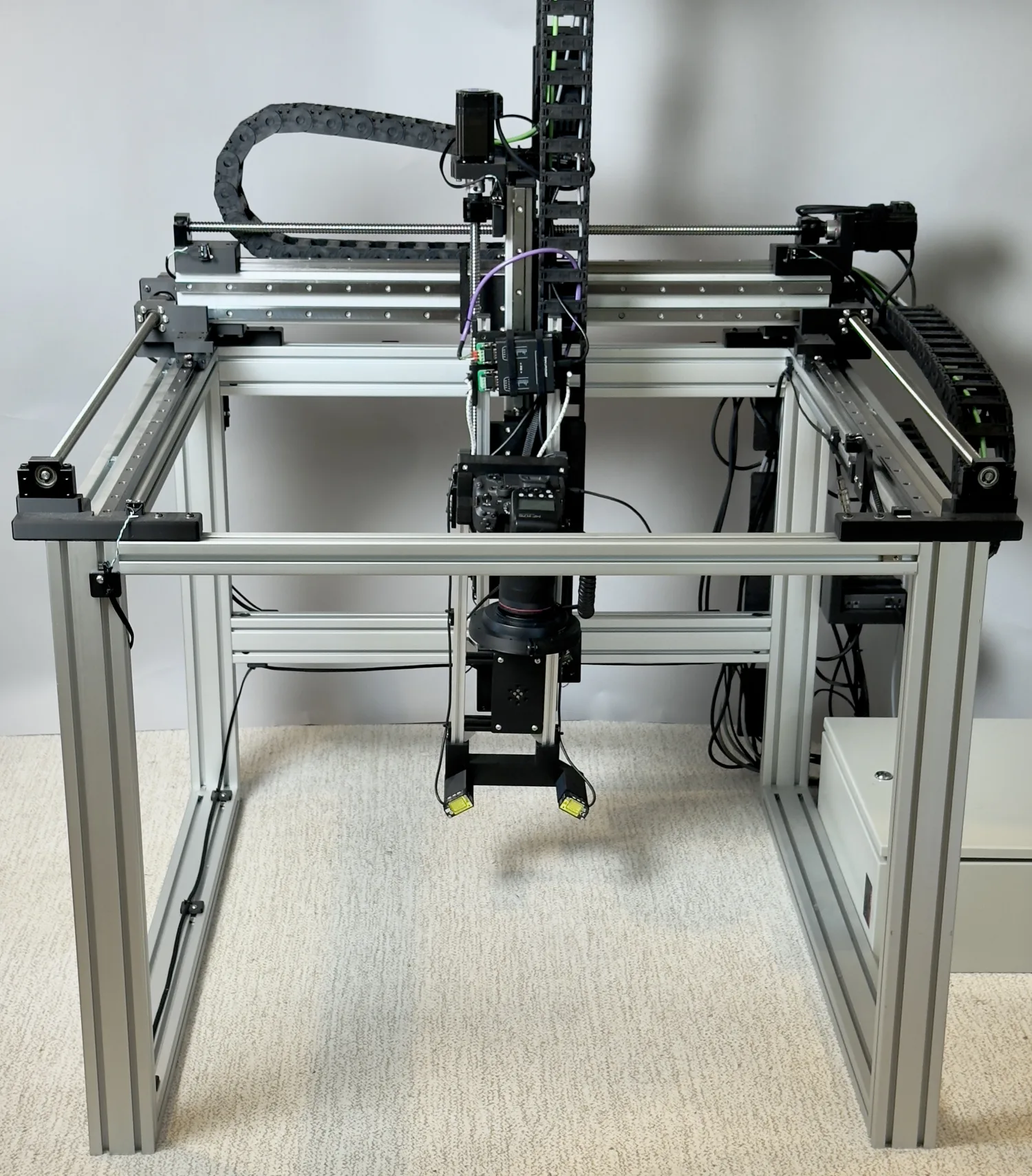
OpenDerm is built primarily from off-the-shelf parts. The video below shows the assembled prototype running a capture sequence at 20× speed.
The gantry moves the camera through four degrees of freedom. Three linear axes position the sensor head within the workspace: X travels along the side rails, Y moves across the top beam, and Z sets the camera height. A rotary axis (RX) tilts the sensor head to align the camera with the skin surface.
X Long travelY Cross travelZ Vertical travelRX Pan rotationOpenDerm · 4-DOF gantry
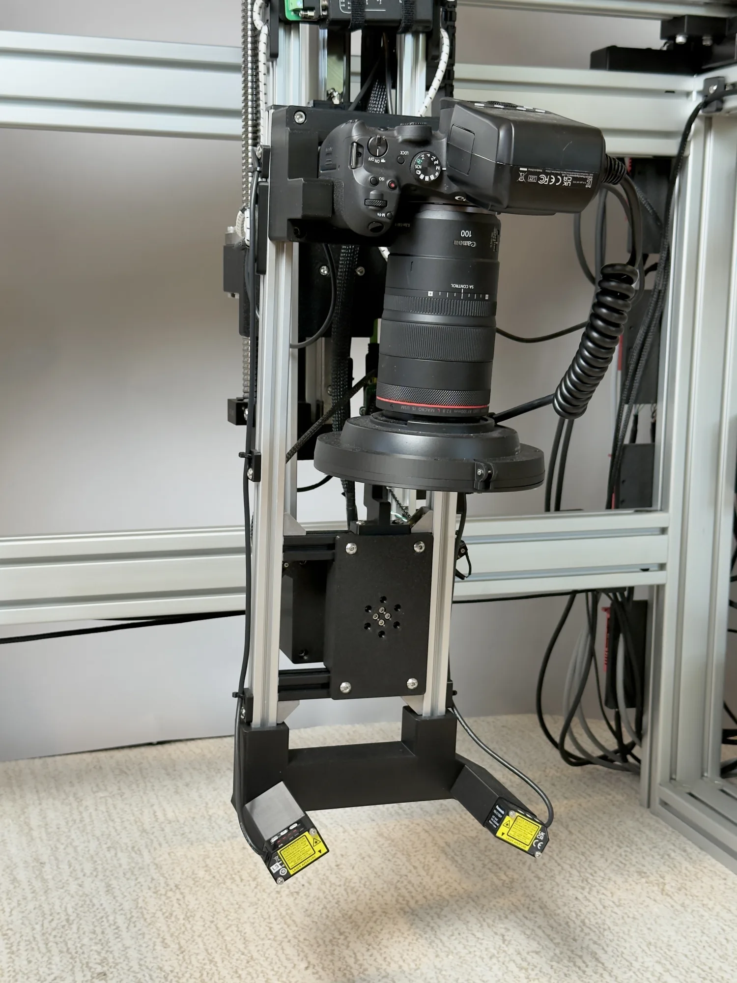
The sensor head carries a Canon EOS R7 with an RF 100 mm macro lens, a Godox MF-R76 ring flash fitted with a cross-polarization filter that reduces specular glare, and two downward-facing laser distance sensors. The distance measurements allow the control system to maintain a consistent working distance and camera orientation relative to the skin.
Canon EOS R7Canon RF 100mm lensGodox MF-R76 Flash +Cross Polarization Filter×2 laser distance sensorsOpenDerm · sensor head
03 / THE SCANS
High-resolution skin imaging
5 mm
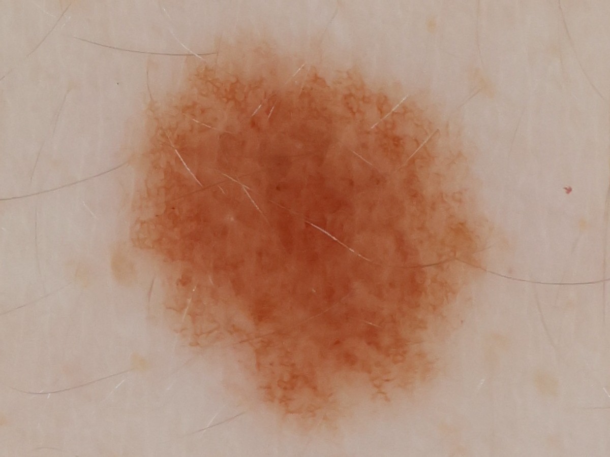
OpenDerm captures skin at 78 pixels per millimeter, resolving details such as a mole’s pigment network and nearby individual hairs. In the interactive viewer, you can explore two registered 3D scans and inspect the lesions measured and matched across them.
At each imaging station, the robot uses two laser sensors to control the camera’s angle and working distance before capturing a high-resolution photograph. The complete pipeline then aligns the overlapping images and reconstructs them as one continuous surface.
Total-body photography remains an emerging field, with limited clinical availability in the United States and many systems still experimental or under development. Three main approaches have emerged for imaging large areas of skin: wide-field total-body photography, close-range robotic scanning, and guided smartphone imaging. Each involves different trade-offs in speed, detail, consistency, footprint, and cost.
OpenDerm captures skin at 78 pixels per millimeter using less than $8,500 in hardware. It is fully open source and provides some of the highest-resolution imaging available at a fraction of the cost of commercial total-body photography systems.
Approach 01
Wide-field total-body photography
Wide-field systems photograph large areas of the body from a distance, either with an array of fixed cameras or a single camera repositioned automatically between views. They provide standardized whole-body records, but individual lesions may require separate close-up or dermoscopic imaging.
SecondsWide-field detail$$$
Examples: Neko · Canfield VECTRA · DermSpectra · FotoFinder
Wide-field capture · fixed-array example
Approach 02
Close-range robotic scanners
A high-resolution camera mounted on a robotic arm or gantry moves close to the skin and systematically across the body while maintaining a controlled distance and viewing angle. Imaging the skin region by region allows a single camera to capture much finer detail than a distant wide-field system, while requiring less space and camera hardware. The trade-off is speed: the body must be scanned sequentially rather than all at once.
MinutesHigh-resolution detail$$
Examples: SquareMind · iToBoS · OpenDerm
Robotic scanner · one moving camera
Approach 03
Guided smartphone imaging
A smartphone application guides a patient, caregiver, or clinician through a standardized series of photographs. This approach requires little dedicated hardware and can be deployed almost anywhere. Its main limitations are image resolution and consistency: lighting, distance, pose, framing, and focus can vary between scans, making subtle longitudinal changes more difficult to measure reliably.
ManualVariable detailNo dedicated hardware$
Examples: SkinIO · MoleMap · Miiskin
Phone & app · guided capture
05 / BUILD IT YOURSELF
Open-source resources for building OpenDerm
The complete bill of materials, CAD assembly, electrical and safety schematics, and step-by-step build instructions are publicly available.